High-Resolution, Fast, and Accurate Imaging and Observation of Live Intracellular Processes and StructuresFLUOVIEW FV3000 Series of Confocal Laser Scanning Microscopes
April 5, 2016

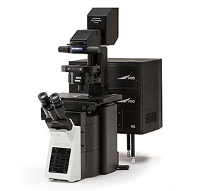
FV3000 confocal laser scanning microscope
Olympus Corporation (President: Hiroyuki Sasa) today announced the global launch, by its Scientific Solutions Business, commencing in July 2016, of the FLUOVIEW FV3000 and FV3000RS. Two new models of confocal laser scanning microscopes provide high-resolution images for fast, accurate imaging of biological samples.
Confocal laser scanning microscopes are used for fluorescent observation*1 of fine structure and movements inside tissue or living cells. These microscopes obtain high-contrast three-dimensional images of microscopic structures inside cells by detecting the small amounts of fluorescent light emitted by a sample as it is scanned by a laser beam. Olympus markets its confocal laser scanning microscopes and multiphoton laser scanning microscopes*2 as the FLUOVIEW series.
The new FV3000 series is the successor to the FV1200. In addition to imaging reactions in cells at high speed and obtaining high-resolution three-dimensional images, the new models have high sensitivity and can capture light even from dim samples. They also support precision observation, including a highly efficient spectral detector that can discriminate fluorescence by wavelength as well as a timing management system that can reliably execute observation schedules that follow a variety of different applications. These features provide faster and more accurate imaging of movements inside tissues or living cells, giving the microscopes a role at the leading edge of research in such fields as the study of cancer biology or industrial and research utility for the development of iPS and other pluripotent cells.
*1 A technique for cell observation that detects small amounts of fluorescent light emitted from fluorescently labeled cells when they are illuminated with an excitation beam.
*2 A microscope that can observe fluorescence from deep inside tissue when a sample is illuminated with a near-infrared laser.
Launch Overview
| Name | Launch Date |
|---|---|
| FV3000 and FV3000RS confocal laser scanning microscopes | July 2016 |
Main Features
- 1. Fast and accurate imaging of biological dynamics using a high-speed scanner*3
- 2. Bright fluorescent imaging of even dim samples made possible by a highly efficient spectroscopic mechanism and high-sensitivity detector
- 3. Improved observation reliability achieved by precise spectroscopy and newly developed time management system
*3 Only available with the FV3000RS confocal laser scanning microscope.
Launch Background
The use of live tissue or cells in an effort to understand their roles and functions is common in basic life science research, drug development, and medical research. Confocal laser scanning microscopes and multiphoton laser scanning microscopes can acquire information from deep within cells or tissues, something that is difficult to achieve with conventional microscopes, and they can be used to perform three-dimensional observation of tiny intracellular structures. As a result, laser confocal microscopes are used at numerous research institutions.
Research applications include cellular biology, stem cells, studies on the commercialization of regenerative medicine, the study of cancer mechanisms, and the development of new medications. This type of research work has a need for faster and more accurate imaging of things like movement inside tissue or intracellular processes.
To meet these research demands, Olympus will launch the new FV3000 series of confocal laser scanning microscopes developed using a combination of optical and digital technologies built up over many years.
Details of Main Features
1. Fast and accurate imaging of biological reactions using a high-speed scanner
The microscopes are fitted with a high-speed scanner capable of capturing images at up to 438 frames per second. This enables the observation of high-speed reactions that take place inside tissue or cells, such as the movement of blood through tissue or intercellular signaling.
2. Bright fluorescent imaging from dim samples made possible by a highly efficient spectroscopic separation mechanism and high-sensitivity detector
The microscopes incorporate a spectroscopic mechanism for the highly efficient discrimination of fluorescence by wavelength and a detector that can capture light with high sensitivity. This enables highly sensitive imaging of the weak amounts of fluorescent light emitted by cells.
3. Improved observation reliability achieved by precise spectroscopy and newly developed time management system
The newly added spectroscopic mechanism enables accurate imaging of intracellular movements by providing precise color separation of regions where fluorescent proteins or dyes overlap. Furthermore, use of the newly developed time management system enables the reliable execution of different observation procedures, including, for example, changing the scanning range or laser light midway through imaging. These features improve the reliability of observation inside tissue or cells.
Main Specifications of FV3000 and FV3000RS
| Laser Light |
405 nm (50 mW), 488 nm (20 mW), 561 nm (20 mW), 640 nm (40 mW)
|
|---|---|
| Scanner |
|
| High Sensitivity-Spectral Detector |
|
| Spectral Detector |
|
| Optional Software |
|
- Olympus's Scientific Solutions Business
- Its main products are optical microscopes and industrial videoscopes and non-destructive testing equipment. Through these products, the Scientific Solutions Business helps to keep the infrastructure of society safe and secure, including research and development in the medical, life science, and industrial fields; quality improvement at production facilities; and inspections of aircraft and other large plant, etc..
Press releases are company announcements that are directed at the news media.
Information posted on this site is current and accurate only at the time of their original publication date, and may now be outdated or inaccurate.
Company names and product names specified are trademarks of their respective owners.

