
Launch of the SpinSR10 Super-Resolution Imaging SystemOptical performance for 120nm resolving power enables live observation of minute changes in subcellular organelles
November 7, 2017
Updated: November 7, 2018
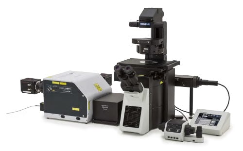
SpinSR10

Left:Confocal Right:Super Resolution
Stress fibers of Hela cell: Antibody staining with Alexa488 (green) for actin, Alexa 568 (red) for myosin heavy chain.
Image courtesy of: Keiju Kamijo,Ph.D. Division of Anatomy and Cell Biology, Faculty of Medicine, TOHOKU Medical and Pharmaceutical University
Olympus Corporation (Representative Director and President: Hiroyuki Sasa) has announced its SpinSR10, the super-resolution imaging system, as a new product in its scientific business. SpinSR10, which allows live observation of intracellular structures and movements such as signal transmission, is to be launched worldwide (with the exception of some regions) from January 2018.
A super-resolution microscope is a microscope with resolving power that surpasses the limits of conventional optical microscopes. The minimum resolving power of general optical microscopes is around 200nm, but a super-resolution microscope has high enough resolving power to observe internal cell structures, which used to be difficult to observe, in fine detail. Therefore, this product is expected to contribute to research in fields such as medicine and biology, which requires observation of fine structures.
With the system’s fast image acquisition speed of 0.005s/frame and 120nm resolving power, the newly-launched SpinSR10 achieves high-speed super-resolution imaging. That imaging can observe detailed changes in subcellular organelles as they happen, which can be expected to contribute to further progress in medical research fields like autophagy*1. This model also features one-touch switching between observation of a wide field of view to super-resolution observation of detailed areas, enabling smooth and convenient observation without wasting time on searching for target areas. The silicone-immersion objectives, designed for observing deep areas, can focus down to the deeper parts of even a thicker sample.
1 One system for breaking down proteins within cells. In 2016 the Nobel Prize in Physiology or Medicine was awarded for research into autophagy.
Launch Overview
| Product name | Launch date |
|---|---|
| SpinSR10 spinning disk-type confocal super-resolution microscope | Scheduled for January 2018 |
Main Features
- Super-resolution imaging at 0.005s/frame enables live observation of minute changes in subcellular organelles
- One-touch switching to super-resolution achieves smooth observation
- The silicone-immersion objectives can observe deep areas, even in thicker samples
Details of Main Features
1. Super-resolution imaging at 0.005s/frame enables live observation of minute changes in subcellular organelles
This model uses Olympus’ unique patented optical technology*2 to attain 120nm resolving power. Applying the spinning disk-type confocal optical system, it enables acquiring live super resolution images at 0.005s/frame. The combination of these two technologies enables rapid super-resolution observation of samples, for live observation of fast, detailed phenomena like autophagy. Therefore, this product is expected to help to further research in medical fields such as cancer and Alzheimer’s disease.
2 Olympus’ unique patented optical technology (patent No.5412394).
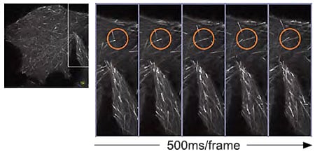
GFP-EB3 at the tip of elongating microtubules in HeLa cell.
Image courtesy of: Dr. Kaoru Katoh ,Biomedical Research Institute, National Institute of Advanced Industrial Sciences and Technology
2. One-touch switching to super-resolution achieves smooth observation
Other than super-resolution observation, this model also supports fluorescence microscopy with a wide field of view, and confocal observation, with one-touch switching between these three modes. After observation of a wide field of view is used to find the target area, the user can easily switch to confocal, and super-resolution observation, a smooth process which makes it quick and easy to find the region of interest.
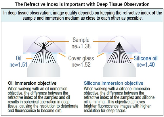
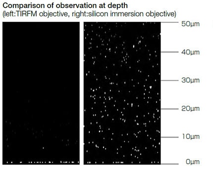
3. The silicone-immersion objectives can observe deep areas, even in thicker samples
Olympus silicone-immersion objectives are designed for deep tissue observation. Observation depth is negatively impacted by spherical aberration caused by refractive index mismatch. The refractive index of silicone oil (ne=1.40) is close to that of living cells or cultured tissue slices (ne=1.38), enabling super resolution imaging of internal cellular structures at tens of micrometers in depth with minimal spherical aberration.
Main Specifications of the SpinSR10
| XY resolving power | 120nm |
| Image acquisition speed (max) | 0.005s/frame |
| Scanning method | Spinning disk type |
| Mounted lasers | Up to 6 colors (2 colors can be used simultaneously for imaging) |
For more information
Press releases are company announcements that are directed at the news media.
Information posted on this site is current and accurate only at the time of their original publication date, and may now be outdated or inaccurate.