
Frame Design to Secure Working Space Olympus Launches Two Types of Upright FV3000 Confocal Laser Scanning MicroscopeAble to observe super-fast phenomena at the industry’s fastest imaging speed of 438 images per second
November 9, 2017
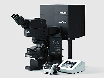
FV3000 Upright Confocal Laser Scanning Microscopes, combination for fixed specimens
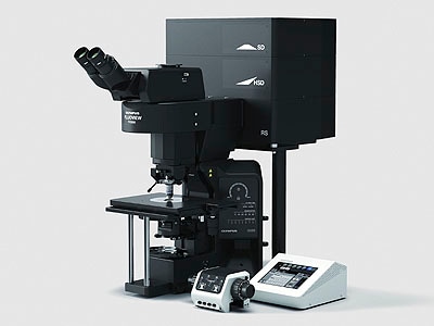
FV3000 Upright Confocal Laser Scanning Microscopes, combination for electrophysiology
Olympus Corporation (Representative Director and President: Hiroyuki Sasa) has announced Two Types of Upright FV3000 Confocal Laser Scanning Microscope, as new products in its scientific business. The new models, which are able to observe phenomena such as super-fast signal propagation in the nervous system, will be launched worldwide in January 2018.
A confocal laser scanning microscope uses lasers to scan a sample stained with fluorochrome, and detects the weak fluorescence emitted by the sample. This method allows observation of fine structures within cells as detailed 3D images. We provide the FV3000 Series of confocal laser scanning microscopes to meet diverse needs.
The two types of upright FV3000 confocal laser scanning microscopes that we are now launching cover the two applications of fixed specimens and electrophysiology. In the configuration for electrophysiology, the frame design provides more working space around the objectives, so that the patch clamp method*1 can be easily operated to observe signal propagation in nerves. By achieving the industry’s fastest imaging speed*2 of 438 images per second, these instruments can capture the super-fast phenomena which accompany signal propagation in nerves. The FV3000 Series supports objectives with a wide range of magnification levels, to provide stepped coverage ranging from macro observation of a wide field of view to micro super-resolution observation.
The addition of these models to the FV3000 lineup enables the series go further, newly covering the field of neuroscience as well as cancer research, stem cell research, and cellular biology.
1 A method that uses fine electrodes to directly measure the propagation activity of electrical signals in nerve cells.
2 As investigated by Olympus. A comparison of four major companies.
Launch Overview
| Product name | Launch date |
|---|---|
| Two Types of Upright FV3000 Confocal Laser Scanning Microscope | Scheduled for January 2018 |
Main Features
- Frame design optimized for electrophysiology experiments, to secure sufficient working space
- Able to observe super-fast phenomena at a maximum speed of 438 images per second
- Supports wide-ranging observation, from macro-scale field of view of 1cm2 down to micro-scale observation of sub-micron sizes
Sales Background
In life sciences and medical fields, living tissues and cells are used in attempts to elucidate their roles and functions. A confocal laser scanning microscope can obtain information in the depth direction, which is difficult with general microscopes, observing detailed intra-cellular structures three-dimensionally. They are chosen by many researchers for that characteristic. Within that context, these microscopes are used in the stem cell and cancer research fields for practical development of regenerative medicine, to elucidate the mechanisms of cancer, and in work to develop therapeutic drugs. Such research demands an accurate and fast grasp of reactions within living tissue and cells. We provide the FV3000 Series of confocal laser scanning microscopes to meet such needs. We have now expanded that lineup by launching two types of upright microscope, the combination for fixed specimens, and the combination for electrophysiology. They expand the reach of the series to cover neuroscience in addition to previously covered fields.
Details of Main Features
1. Frame design optimized for electrophysiology experiments, to secure sufficient working space
Electrophysiology experiments observe reactions and phenomena which occur in nerves and cells when current flows in a sample, in order to elucidate their mechanisms. To that end, the large working space design provides ample room around the objective to easily set up electrophysiology equipment.
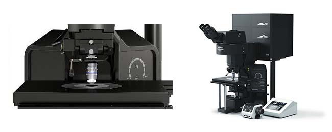
The frame design of the configuration for electrophysiology
2. Able to observe super-fast phenomena at a maximum speed of 438 images per second
This model is equipped with a high-speed scanner to reach 438 frames per second, the fastest imaging speed in the industry. That enables observation of super-fast phenomena in the neuroscience field. Research in this field can capture super-fast changes in detail, happening within one second as nerve cells transmit signals to each other.
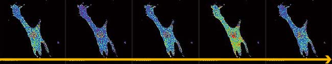
IMD ratio images of spontaneous Ca2+ oscillation in a beating rat cardiomyocyte expressing yellow cameleon.
Image data courtesy of Dr. Yusuke Niino and Dr. Atsushi Miyawaki,Cell Function Dynamics, Brain Science Institute of RIKEN.
3. Supports wide-ranging observation, from macro-scale field of view of 1cm2 down to micro-scale observation of sub-micron sizes
The FV3000 Series supports objectives with magnification levels ranging from 1.25X to 150X, to provide stepped coverage ranging extensively from macro observation of a wide field of view to micro super-resolution observation. To that end, these products can be used to check the retina sample in its entirety, as shown below, then narrow in on the region of interest for micro-scale observation of the linkages of individual nerve cells.
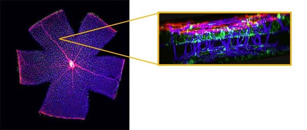
Mouse retina
One shot overview image acquired by PLAPON2X and 3D image acquired by UPLSAPO40XS silicone immersion objective.
Image data courtesy of Dr. Yoshiaki Kubota, The Laboratory of Vascular Biology Center for Integrated Medical Research, School of Medicine, Keio University.
For more information
Press releases are company announcements that are directed at the news media.
Information posted on this site is current and accurate only at the time of their original publication date, and may now be outdated or inaccurate.