
Maximize Resolution in Deep Imaging for Neuroscience Research
Olympus Launches TruResolution Objectives for Multiphoton Laser Scanning Microscopes
Industry-First Automatic Spherical Aberration Compensation Function Developed Jointly with RIKEN
January 17, 2018
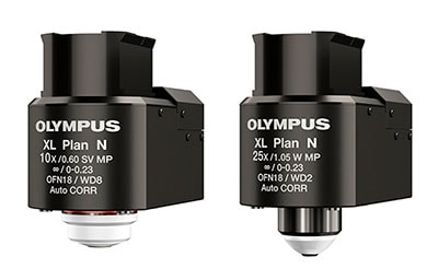
FV30-AC25W and FV30-AC10SV Objectives with Automatic Spherical Aberration Compensation
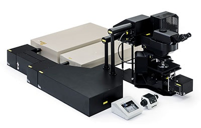
FLUOVIEW FVMPE-RS Microscope
Olympus Corporation (Representative Director and President: Hiroyuki Sasa) today announced the joint development with the RIKEN Brain Science Institute of the FV30-AC25W and FV30-AC10SV objectives for multiphoton laser microscopes, featuring automatic spherical aberration compensation. These new products from Olympus’ Scientific Solutions business will be launched worldwide on January 17, 2018.
Neuroscience research needs precise three-dimensional imaging of rapid biological processes, such as nerve excitation and conduction or synaptic transmission. Olympus’ proven FLUOVIEW FVMPE-RS multiphoton laser scanning microscope facilitates such research. This high-performance model of the FLUOVIEW microscope series enables deep imaging, an important requirement in neuroscience.
The new FV30-AC25W and FV30-AC10SV objectives were designed specifically for the multimode FLUOVIEW FVMPE-RS microscope and are equipped with motorized correction collars that can automatically and dynamically compensate for spherical aberration*1 while maintaining the focus position. This capability enables clear and bright imaging, even for the fine structure of nerve cells. These new objectives were developed based on research by the RIKEN BSI-Olympus Collaboration Center*2.
1 Focal distortion caused by the different refractive indices of the sample and immersion liquid.
2 Joint research facility of Olympus and the RIKEN Brain Science Institute in Japan.
Launch Overview
| Name | Launch Data |
|---|---|
| FV30-AC25W and FV30-AC10SV Objectives with Automatic Spherical Aberration Compensation | January 17, 2018 |
Main Features
- Automatic spherical aberration compensation enables bright and clear deep imaging.
- Optimal compensation is easily achievable with the assistance of dedicated software.
Launch Background
Medical and life science research examines living tissue or cells to elucidate their purpose and functions. In neuroscience, fast and precise imaging of phenomena such as nerve cell excitation and conduction or synaptic transmission, both at the surface and in deeper regions of the brain, has an important role in helping determine mechanisms. Olympus’ FLUOVIEW FVMPE RS multiphoton laser scanning microscope meets the needs of users engaged in such research.
A problem with deep imaging in brain science research is that spherical aberration increases when imaging deeper locations, making it more difficult to obtain clear images. Spherical aberration is caused by the different refractive indices of the sample and the liquid in which it has been immersed for imaging. The new objectives equipped with mechanisms for automatic compensation of spherical aberration overcome this problem. These objectives are the result of combining Olympus’ knowledge of optics with the specialist expertise in neuroscience possessed by the RIKEN Brain Science Institute (RIKEN BSI).
The new objectives emerged from work undertaken by the RIKEN BSI-Olympus Collaboration Center to identify the numerous issues that arise in the process of supporting observational experiments by researchers.
Details of the Main Features
1. Automatic spherical aberration compensation enables bright and clear deep imaging.
In the past, compensating spherical aberration when using the objectives on Olympus microscopes required manual adjustments (rotating a correction collar*3 attached to the objectives), which were difficult to make without shifting the focus location. These new TruResolution objectives automatically compensate for spherical aberration through an algorithm that finds the best collar setting at each depth using a motorized correction collar. Although spherical aberration increases when imaging deeper into the samples, TruResolution objectives maintain optimal compensation as the depth changes. The result is consistently bright and clear images.
3 A ring-shaped mechanism that moves the internal objectives to compensate for spherical aberration.
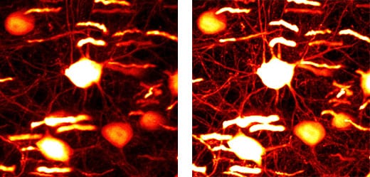
Image of a nerve cell in the brain (depth: 600 µm, without compensation [left], with compensation [right])
2. Optimal compensation is easily achievable with the assistance of dedicated software.
The software was developed by combining RIKEN’s expertise in neuroscience with Olympus’ knowledge of optics. The optimal compensation can be applied automatically simply by having the software scan the sample a number of times before performing actual measurements to calculate the spherical aberration.
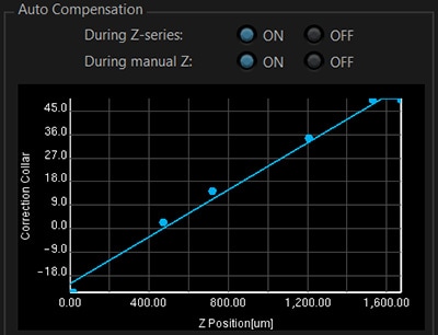
Interface for automatic spherical aberration correction
Graph of correction value calculated automatically from scan results
Main Specifications
| Name | FV30-AC10SV | FV30-AC25W |
|---|---|---|
| Magnification | 10 | 25 |
| Numerical Aperture (NA) | 0.6 | 1.05 |
| Working Distance (W.D.) | 8mm | 25mm |
| Immersion Liquid | SCALEVIEW-A2*4 (Water, silicone oil and normal oil available) |
Water |
4 An optical clearing agent that enables deep imaging of sample tissue without damaging it.
For more information
Press releases are company announcements that are directed at the news media.
Information posted on this site is current and accurate only at the time of their original publication date, and may now be outdated or inaccurate.