
High-Sensitivity Model of the SpinSR10 Super Resolution Imaging System
Delivers Clearer Images and Three Times the Brightness of the Standard Model
November 1, 2018
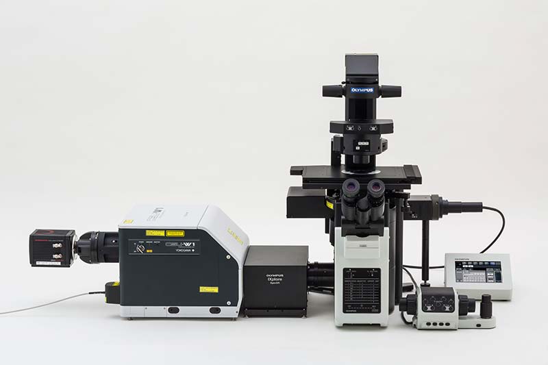
High-sensitivity model of the SpinSR10 super resolution imaging system
Olympus Corporation (Representative Director and President: Hiroyuki Sasa) today announced the worldwide launch (certain regions excluded), commencing in December 2018, of a new product from its scientific solutions business: the high-sensitivity model of the SpinSR10 super resolution imaging system that offers three times the brightness of the standard model.
Super resolution microscopes have a resolving power that surpasses the limits of conventional optical microscopes. The minimum resolving power of a standard optical microscope is about 200 nm, but a super resolution microscope has the resolving power to observe internal cell structures in fine detail. The high-sensitivity SpinSR10 microscope will contribute to medical and biological research where observing fine structures is critical.
The high-sensitivity SpinSR10 microscope combines fast 0.005 seconds/frame acquisition speeds and 120 nm super resolution with three times the brightness of the standard model. The new scanner unit has upgraded optics to acquire images with greater clarity than before. In addition, the increased brightness enables users to capture images with the same brightness as the previous model using less laser light, helping reduce damage to the sample or fading fluorescent dyes. This is especially useful during long-term observations. With these features, this new model will contribute to medical research in cancer, neurology and other fields.
Launch Overview
| Name | Launch Data |
|---|---|
| SpinSR10 Spinning Disk Confocal Super-Resolution Microscope High-Sensitivity Model | December 2018 |
Main Features
- Improved brightness (three times that of the standard model) provides clearer images
- Reduces sample damage and the fading of fluorescent dyes due to laser light
Details of Main Features
1. Improved brightness (three times that of the standard model) provides clearer images
The high-sensitivity SpinSR10 model has three times the brightness of the standard model thanks to enhancements to the disk used for fluorescent light detection in the scanner unit. The new model has the same fast image acquisition speed (up to 0.005 s/frame) and resolution (120 nm) as the standard model, enabling high-speed imaging of the small internal structures of cells and any changes they undergo. Furthermore, the high-sensitivity model provides greater image clarity than the standard model when viewing samples under the same conditions.
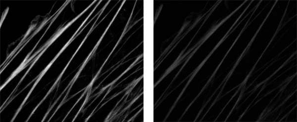
Brightness comparison
Left: High-sensitivity model, Right: Standard model
Images of the Ptk2 cell acquired under the same conditions (Objectives: UPLSAPO100XS).
Image clarity is shown on the magnified section.
2. Reduces sample damage and the fading of fluorescent dyes due to laser light
The improved brightness means that users can acquire images with the same brightness as the standard model using a lower laser output, helping reduce damage to the sample and fading fluorescent dyes caused by the laser light. As a result, it is possible to observe changes in internal cell structures over long periods of time, even when observing easily damaged live cell samples. This provides highly reliable super resolution live cell imaging, more accurate data, and reduced damage to cells.
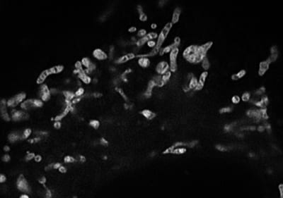
Real-time live cell imaging of mitochondria (10FPS)
Image data courtesy of Kaoru Kato, PhD, National Institute of Advanced Industrial Science and Technology Biomedical Research Institute
Acquired with CSU-W1 SoRa (manufactured by Yokogawa Electric Corporation) incorporated in the high-sensitivity SpinSR10 microscope
Performance of the standard model of the SpinSR10 super resolution imaging system
For more information
Press releases are company announcements that are directed at the news media.
Information posted on this site is current and accurate only at the time of their original publication date, and may now be outdated or inaccurate.